
Need to communicate at AMA: Power 2025 or AMA: Automotive & Mobility 2025? Submit your software now!
Researchers from the Royal School of Surgeons in Eire (RCSI) College of Medication and Well being Sciences and Trinity School Dublin have developed a 3D printed implant which will help the restore of spinal twine accidents by delivering electrical stimulation to broken nerve tissue.
Led by Dr Ian Woods and Professor Fergal O’Brien at RCSI’s Tissue Engineering Analysis Group (TERG), in partnership with the AMBER Centre at Trinity School Dublin, the research was revealed in Superior Science. It was supported by the Irish Rugby Soccer Union Charitable Belief (IRFU-CT), and the Irish Analysis Council.
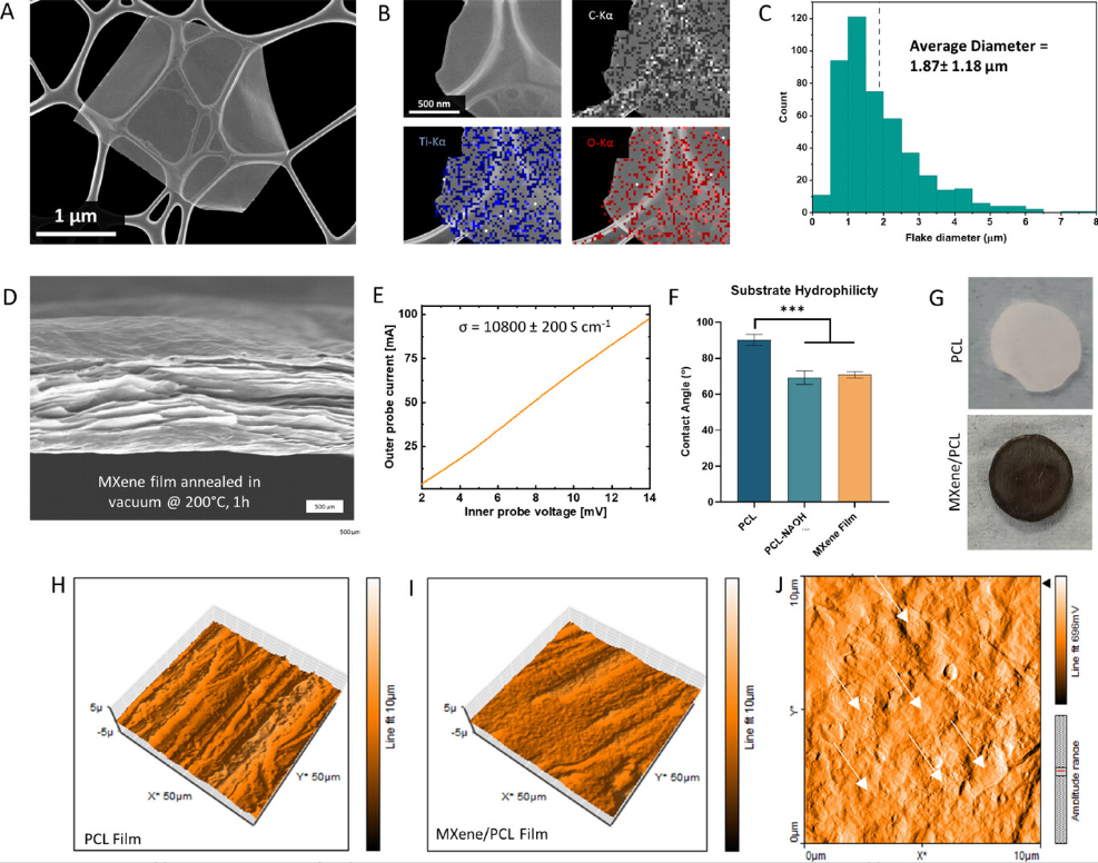
The implant is produced from a comfortable, gel-like extracellular matrix composed of hyaluronic acid, collagen type-IV, and fibronectin, designed to resemble the pure atmosphere of the spinal twine. Inside this matrix, the group inserted a nice mesh manufactured from plastic fibers (polycaprolactone, or PCL) coated with MXene nanosheets, microscopic flakes of a conductive materials that assist transmit electrical indicators with out harming surrounding cells.
“Selling the regrowth of neurons after spinal twine damage has been traditionally troublesome; nonetheless our group is creating electrically conductive biomaterials that would channel electrical stimulation throughout the damage, serving to the physique to restore the broken tissue,” explains Professor O’Brien.
Printed fibers information electrical therapeutic
Utilizing a method referred to as soften electrowriting, the researchers 3D printed these fibers with excessive precision, spacing them at completely different densities. They created three variations of the implant: low-, medium-, and high-density fiber networks, with spacing starting from 1000 to 500 µm. Every variation produced completely different ranges {of electrical} conductivity, starting from 0.081 to 18.87 siemens per meter (S/m).
When freeze-dried, the scaffold retained porosity better than 99% and maintained a compressive stiffness between 0.6 and three.25 kPa, much like the softness of spinal twine tissue. To check effectiveness, the researchers grew human-derived neurons, astrocytes, and microglia on the implant.
Neurons confirmed considerably elevated development and metabolic exercise on the MXene-coated fibers in comparison with controls. Astrocytes had been much less reactive, which is favorable for nerve restore, and microglia confirmed no indicators of irritation. These outcomes confirmed that the implant was each protected and appropriate with central nervous system cell sorts.
Additional exams assessed the implant’s efficiency beneath electrical stimulation. In a single experiment, neurons had been cultured on the implant for seven days. These on high-density scaffolds and uncovered to electrical indicators grew axons averaging 108.5 µm, in comparison with 74.3 µm in unstimulated controls and 67.4 µm on unstimulated MXene scaffolds. Medium-density scaffolds, nonetheless, yielded the longest neurites per cell and the best ranges of BII-tubulin, a marker of neuron maturity.
In a extra superior mannequin, the group used neurospheres, i.e., 3D clusters of neural stem cells from the olfactory bulb of mice. These cells can change into numerous mind and nerve cell sorts. When stimulated electrically on the implant, these on medium- and high-density scaffolds developed longer axons and confirmed extra indicators of maturing into neurons. Axon lengths reached 203.6 µm within the high-density group, in comparison with 94.1 µm on non-conductive controls and 88.6 µm within the low-density group.
The construction and spacing of the conductive fibers considerably influenced the affect {of electrical} stimulation. Whereas a tighter mesh improved sign supply, a medium-density design supplied the very best atmosphere for general cell development. MXene content material remained beneath 0.3% of the scaffold quantity, but proved extremely efficient when strategically organized.
The venture additionally benefited from insights offered by an advisory group that included clinicians, researchers, and critically injured rugby gamers. Supported by (IRFU-CT), the group helped researchers perceive the real-world challenges of spinal twine damage and offered steerage on affected person priorities. As Dr. Woods places it, “our common conferences allowed for a constant change of enter, concepts and outcomes.”
Whereas nonetheless in early improvement, the implant gives a brand new method to combining electrical stimulation with comfortable, biocompatible supplies in a exactly tunable 3D printed format.

3D printed implants to revive spinal twine perform
Like different areas of regenerative drugs, 3D printing gives robust potential to enhance outcomes and enhance the effectiveness of spinal restore procedures.
Israeli regenerative drugs agency Matricelf examined a bioprinted spinal twine implant developed utilizing know-how from Tel Aviv College (TAU) on paralyzed mice with notable outcomes. Researchers started by reprogramming cells from a affected person’s stomach fats into stem cells, then embedded them in a customized hydrogel produced from the affected person’s personal extracellular matrix.
These had been bioprinted into spinal cord-like neuronal networks designed to keep away from immune rejection. After implantation, all mice with acute paralysis regained motion, whereas 80% of chronically paralyzed mice recovered. The tactic mimicked embryonic spinal improvement, and Matricelf sought to start human trials by late 2024 following additional security research.
Moreover, researchers from the College of California San Diego’s College of Medication and Institute of Engineering in Medication (IEM) developed a 3D printed spinal twine implant that restored motor perform in rats with extreme accidents. Every 2 mm implant was produced in simply 1.6 seconds, that includes 200 µm-wide channels that guided neural stem cell development and helped reconnect severed axons.
Implanted into damage websites, the scaffold supported pure vascularization and enabled vital hind limb restoration. To reveal medical potential, the group additionally printed four-centimeter human-scale implants from MRI scans in beneath 10 minutes, shifting nearer to future human trials.
What 3D printing traits do you have to be careful for in 2025?
How is the way forward for 3D printing shaping up?
To remain updated with the newest 3D printing information, don’t overlook to subscribe to the 3D Printing Trade e-newsletter or observe us on Twitter, or like our web page on Fb.
Whilst you’re right here, why not subscribe to our Youtube channel? That includes dialogue, debriefs, video shorts, and webinar replays.
Featured picture exhibits the Impact of MXene-ECM Scaffold Conductivity and 3D printed Micro-Mesh Design on Neuronal Cell Habits in Response to Steady Electrical stimulation. Picture by way of RCSI.
