
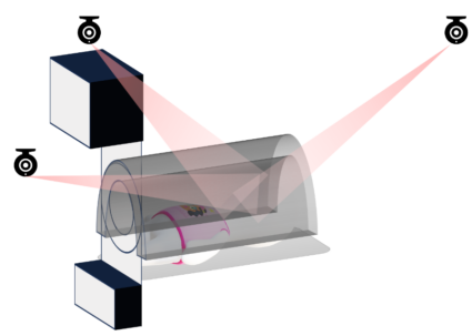
 Illustration of the radiotherapy room and the occlusion downside confronted by ceiling-mounted cameras on this software.
What was the subject of your PhD analysis and why was it an attention-grabbing space?
My matter of analysis was creating an optical tactile sensor to trace head movement throughout radiotherapy. I labored on each the {hardware} and software program growth of this sensor, although my focus was totally on the software program facet. Its significance comes from the truth that throughout radiotherapy, sufferers present process head and neck most cancers therapy are sometimes immobilised. That is normally accomplished utilizing a thermoplastic masks, which might really feel very claustrophobic, or a stereotactic body. Frames are extra frequent for mind cancers, however they must be surgically inserted into the affected person’s cranium utilizing pins. Both of those immobilisation instruments could also be used relying on the state of affairs. When sufferers are uncomfortable, they’re extra prone to transfer, which impacts the accuracy of therapy, particularly with thermoplastic masks.
One other main concern is that present methods use ceiling-mounted cameras to file affected person movement. These cameras can’t be positioned too near the affected person due to the electromagnetic atmosphere across the gear. Their view can be regularly occluded as a result of the affected person strikes right into a tunnel to obtain the ionising beams, which makes it troublesome to seize rotational movement.
One various is an infrared digicam with a nostril marker, however this solely captures translational movement. At the moment, when a nostril tracker detects motion past a sure threshold, therapy is paused, the affected person is repositioned, and therapy resumes. It’s troublesome to adapt this technique to reliably measure the rotational movement of the affected person’s head within the radiotherapy atmosphere.
That is the place the Movement Seize Pillow (MCP) is available in, which comprises the optical tactile sensor I developed. The objective with this technique is just like the nostril tracker, however with extra correct rotational suggestions for the radiographer. It may be positioned beneath the affected person’s head and connected to the therapy mattress. It estimates how a lot the affected person’s neck is rotating and improves affected person consolation. Radiographers can obtain real-time suggestions on each translational and rotational motion. The benefits of this technique are that there are not any occlusions, as a result of the pillow is in direct contact with the affected person’s head, and it’s extra appropriate with radiotherapy environments as a result of the sensor is non-ferromagnetic. Its premise is to keep up affected person consolation, keep compact and straightforward to combine into the pre-existing methods for radiotherapy, while bettering the accuracy of the therapy by real-time head monitoring.
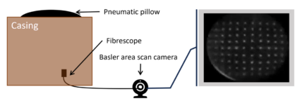 Labelled diagram of the Movement Seize Pillow – Optical tactile sensor for head monitoring throughout radiotherapy. The pneumatic pillow is a deformable rubber-like sheet with embedded white markers, held in its convex form utilizing air strain. The fibrescope represents a non-ferromagnetic fibre optic bundle used as a lens extension to an space scan digicam. The digicam is ferromagnetic and would require secure positioning and fixation.
What have been the principle contributions of your work?
There have been 4 primary contributions to my work. The first contribution centered on making the system extra non-ferromagnetic and bettering the imaging and monitoring method. Earlier work used a webcam and binary picture processing throughout the optical tactile sensor to trace marker displacement. I in the end determined to make use of a fibrescope, optical circulation monitoring algorithm, and grayscale imaging as a substitute, which improved the sensor’s monitoring potential.
The second contribution centered on optimising marker density. The optical tactile sensor consists of an array of markers on a deformable rubber-like sheet, resembling a pillow. The deformation of those markers is captured by the digicam. I investigated how dense the marker array wanted to be by adjusting the spacing between markers to find out what labored greatest for this software.
The third contribution concerned sensor fusion to enhance reliability and robustness. To do that, I built-in a gyroscope and used Kalman filtering to fuse information from the gyroscope and the MCP. This was vital for Gamma Knife methods, that are radiosurgery platforms used for mind cancers. They have a tendency to have greater accuracy necessities than linear accelerators, that are generally used for head and neck cancers, and decrease constraints on the usage of ferromagnetic parts.
The ultimate contribution was a participatory design research performed in collaboration with clinicians and the social sciences division. We explored how the MCP could possibly be built-in into hospital workflows and assessed its feasibility.
How possible is it to combine this sensor into hospital workflows?
Clinicians did appear to be very on board with it, however the research was extra qualitative than quantitative. Whereas they felt the thought had advantage, there have been reservations about adopting new expertise and the related studying curve.
They have been additionally involved about accuracy. Bettering accuracy and reliability is important for clinicians to really feel assured utilizing the system. At current, additional growth is required earlier than it may be extensively applied.
What future work is deliberate on this space?
One space to research is the variations between the model and participant information. The pillow form is managed by a pneumatic system with a strain sensor and air pump. When the affected person or model strikes, strain modifications happen. The system compensates to keep up a set strain, however this introduces errors within the movement readings. The model produced extra errors than the participant information. It might not precisely simulate human movement on the pillow, and the testing setup might introduce discrepancies that don’t mirror real-world behaviour.
So, future work contains stabilising and refining the strain management system to enhance reliability. If vital, reconsidering the usage of gel on the sensors could possibly be an possibility. Gel had been used beforehand however was deserted because of clinician issues about attenuation of ionising beams. Nonetheless, if avoiding gel considerably compromises sensor efficiency, revisiting this method could also be worthwhile.
As well as, extra participant information assortment is required. Not all beforehand collected information could possibly be used because of ground-truth measurements being partially occluded within the experimental setup. Extra participant research would offer a clearer understanding of efficiency throughout completely different people. One other precedence is bettering the fibrescope’s decision and angle to higher visualise high-density marker arrays. {Hardware} upgrades would assist guarantee a clearer subject of view and enhance general system efficiency.
About Bhoomika
 |
Bhoomika Gandhi is a latest PhD graduate from the College of Sheffield Medical Robotics group. Her undergraduate diploma was in Bioengineering – Medical Gadgets and Devices, with management engineering and robotics being the important thing themes. |
Ella Scallan
is Assistant Editor for AIhub
