
Engineered tissue grafts might assist carry out key liver features and profit hundreds of individuals residing with liver failure.
The liver is likely one of the physique’s hardest-working organs, finishing up a whole lot of important jobs, from filtering toxins and metabolizing medicines to producing proteins important for blood clotting. But when it fails, the one definitive therapy is commonly a transplant, an answer restricted by a continual scarcity of donor organs.
MIT engineers have now developed injectable “mini livers” that, in mice, survived for no less than two months whereas performing most of the features of wholesome liver tissue.
“We consider these as satellite tv for pc livers. If we might ship these cells into the physique, whereas leaving the sick organ in place, that would supply booster perform,” says Sangeeta Bhatia, the John and Dorothy Wilson Professor of Well being Sciences and Know-how and of Electrical Engineering and Pc Science at MIT, and a member of MIT’s Koch Institute for Integrative Most cancers Analysis and the Institute for Medical Engineering and Science (IMES).
Bhatia is the senior creator of the examine, which was printed within the journal Cell Biomaterials. MIT postdoc Vardhman Kumar is the paper’s lead creator.
Restoring liver perform
The liver carries out about 500 important jobs, from serving to management blood clotting to clearing micro organism from the blood and breaking down medicine. Many of those duties rely on hepatocytes, the liver’s predominant useful cells.
For greater than a decade, Bhatia’s lab has been creating methods to revive hepatocyte exercise with out requiring a surgical liver transplant. One technique is to put hepatocytes inside a biomaterial reminiscent of a hydrogel, however that method nonetheless requires surgical procedure to implant the gel.
Injecting hepatocytes straight into the physique might keep away from that surgical procedure. On this examine, Bhatia’s lab aimed to make that method simpler by giving the cells an engineered surroundings that would assist them survive and permit docs to trace graft well being with out one other invasive process.
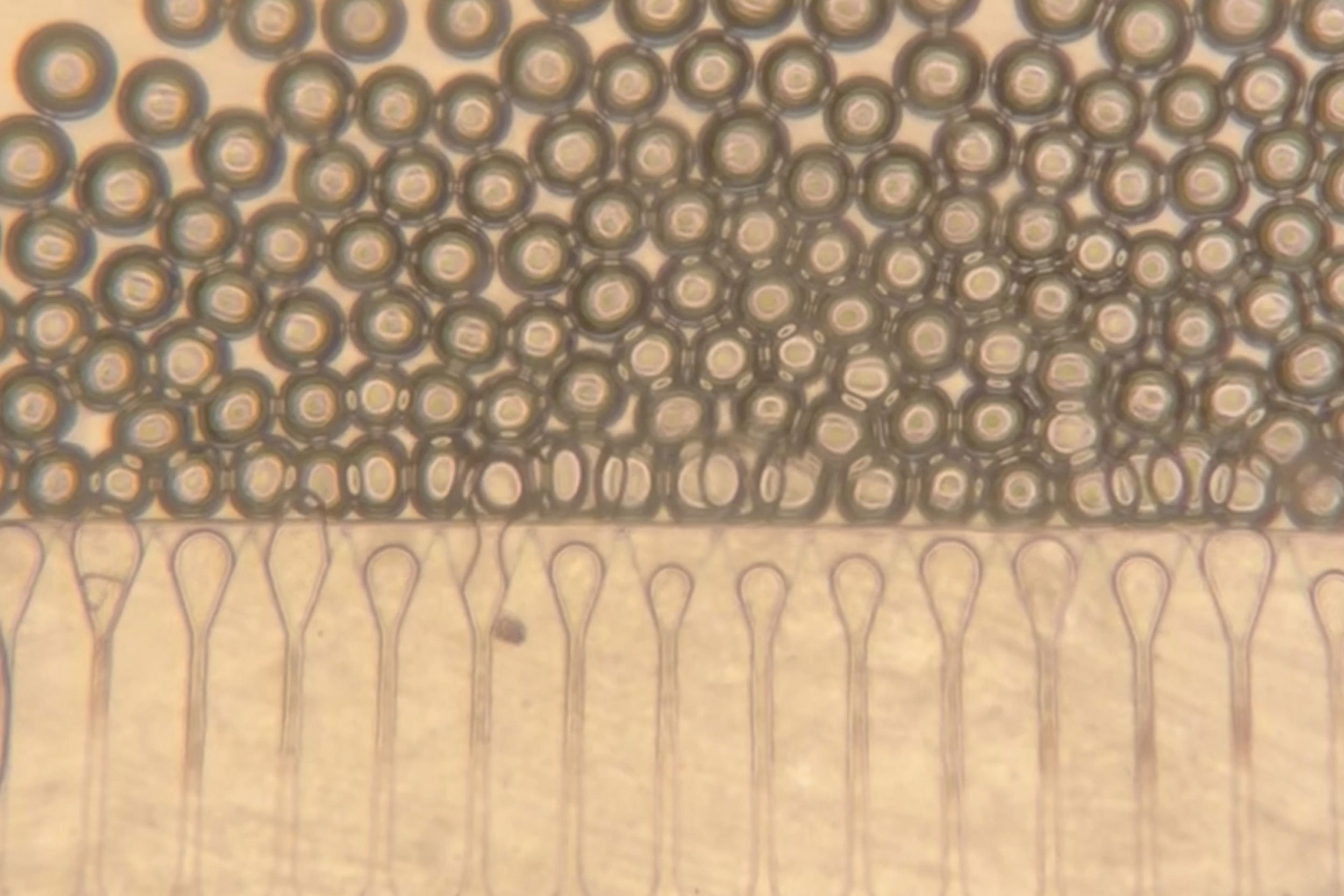
The answer was to inject the cells along with hydrogel microspheres. These tiny spheres assist the cells stay clustered and join with close by blood vessels. They’ll behave like a liquid when packed collectively, which permits them to cross by a syringe, then return to a strong construction as soon as contained in the physique.
In recent times, hydrogel microspheres have been studied as instruments for wound therapeutic as a result of they permit cells to maneuver into the areas between the spheres and kind new tissue. Within the new work, the MIT group tailored the identical fundamental thought to assist hepatocytes construct a secure graft after injection.
“What we did is use this expertise to create an engineered area of interest for cell transplantation,” Kumar says. “If the cells are injected within the absence of those spheres, they’d not combine effectively with the host, however these microspheres present the hepatocytes with a distinct segment the place they’ll keep localized and turn into related to the host circulation a lot quicker.”
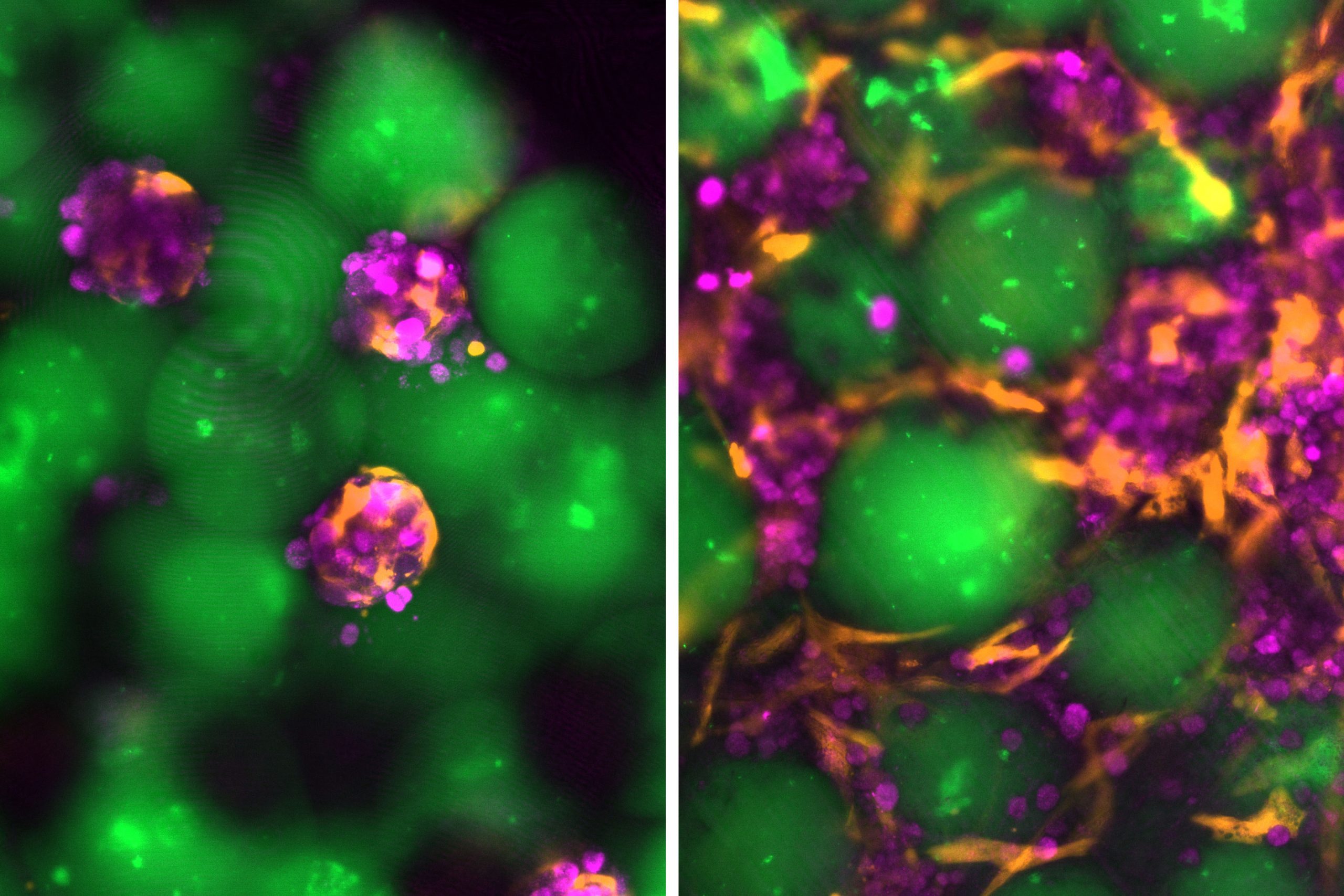
The injected materials additionally incorporates fibroblast cells, which assist hepatocyte survival and encourage blood vessels to develop into the graft.
Working with Nicole Henning, an ultrasound analysis specialist on the Koch Institute, Bhatia’s lab developed an ultrasound-guided syringe technique to put the cell combination within the physique. The identical imaging technique may also be used after injection to comply with the implant’s stability over time.
For this examine, the mini livers have been positioned in fats tissue within the stomach. Future variations might doubtlessly be delivered to different components of the physique, together with the spleen or areas close to the kidneys. If the graft has sufficient room and a powerful blood provide, the injected hepatocytes can act very like hepatocytes contained in the liver.
“For a overwhelming majority of liver issues, the graft doesn’t want to take a seat near the liver,” Kumar says.
A substitute for transplantation
In mouse experiments, the combination of liver cells and microspheres was injected into fatty tissue referred to as perigonadal adipose tissue. After the cells settled in place, they shaped a dense and secure construction. As time handed, new blood vessels grew into the graft, serving to hold the hepatocytes alive and useful.
“The brand new blood vessels shaped proper subsequent to the hepatocytes, which is why they have been capable of survive,” Kumar says. “They have been capable of get the vitamins delivered proper to them, they have been capable of perform the way in which they’re alleged to, and so they produced the proteins that we count on them to.”
The cells remained alive and continued releasing specialised proteins into the animals’ circulation for eight weeks, which was the complete period of the examine. That outcome suggests the method might someday function a long-term therapy for liver illness, in line with Bhatia’s lab.
“The best way we see this expertise is it will probably present a substitute for surgical procedure, however it will probably additionally function a bridge to transplantation the place these grafts can present assist till a donor organ turns into obtainable,” Kumar says. “And if we predict they may want one other remedy or extra grafts, the obstacles to try this are a lot much less with this injectable expertise than present process one other surgical procedure.”
With the present model of the expertise, sufferers would most likely want immunosuppressive medicine. Bhatia’s lab is now finding out attainable methods round that limitation, together with “stealthy” hepatocytes that would keep away from immune assault or hydrogel microspheres that launch immunosuppressants straight on the graft website.
Reference: “Picture-guided injectable area of interest for hepatocyte transplantation” by Vardhman Kumar, Joa Yun, Susanna Okay. Elledge, Nicole Henning, Katarzyna A. Grzelak, Ashley D. Westerfield, Amy Stoddard, Favour A. Oladimeji, Virginia Spanoudaki, Kasturi Chakraborty, Savan Okay. Patel, Heather E. Fleming, Christopher S. Chen and Sangeeta N. Bhatia, 3 March 2026, Cell Biomaterials.
DOI: 10.1016/j.celbio.2026.100378
