
Stanford researchers have developed a microscope that may present how nanostructures work together inside dwelling cells on the highest decision achieved up to now.
The view into dwelling cells simply obtained higher.
Stanford researchers have merged two microscopy strategies to construct a novel instrument that may seize cell buildings interacting in actual time at an unprecedented decision of 120 nanometers. It’s the highest decision but achieved with out fluorescent labels.
The expertise, referred to as Interferometric Picture Scanning Microscopy, or iISM, offers scientists a solution to watch mobile buildings of their broader surroundings, together with how they react to invaders similar to pathogens or to medicine. The advance is described within the journal Gentle: Science and Purposes.
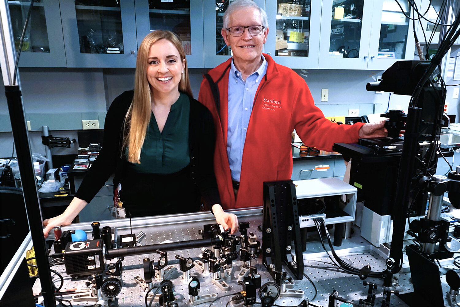
“This new microscope gives a improbable new view into the cell, the place you possibly can see the tiny buildings and machines within the cell transferring, altering, and interacting with out having so as to add fluorescence to watch them,” mentioned senior writer W.E. Moerner, the Harry S. Mosher Professor of Chemistry in Stanford’s College of Humanities and Sciences. “It’s an exquisite look into these complicated little mobile bins that drive our life.”
The talents of iISM may assist new discoveries throughout many areas of the life sciences, together with analysis on illness mechanisms, drug improvement, and interactions between crops and microbes.
Though iISM doesn’t attain the identical decision as some extremely specialised microscopes, its label-free method provides main advantages. Scientists can observe many mobile buildings on the identical time and comply with them for longer intervals. By comparability, fluorescence-based strategies often mark only some chosen buildings directly. Fluorescent indicators also can fade over time. As well as, the labels may be tough to introduce and will generally alter the habits of the buildings being studied.
The iISM additionally works with a lot decrease illumination energy than related excessive distinction label-free strategies. That reduces the possibility of light-related harm in dwelling cells and makes it much less probably that the imaging course of will disturb the small, fragile buildings underneath commentary.
“Each methodology has its benefits and drawbacks, and we consider in a complementary implementation sooner or later,” Kueppers mentioned. “If we use the strengths of fluorescence for molecular specificity and the power of iISM for label-free context and dynamics, we will actually begin tackling questions which have been tough to deal with earlier than.”
Many ‘eyes’ on the identical level
The iISM reaches increased decision and sensitivity by combining the strengths of two microscopy approaches. That mixture displays the experience of the 2 coauthors. Moerner, who acquired the 2014 Nobel Prize in chemistry for his work on super-resolution fluorescence microscopy, recruited Kueppers to Stanford as a result of her doctoral analysis centered on “interferometric scattering microscopy.”
Scattering is the rationale the sky seems blue. When gentle strikes small particles, as daylight does when it passes by means of the environment and encounters mud, water droplets, and different molecules, it adjustments route and scatters. Particles in Earth’s environment scatter brief blue wavelengths extra strongly than pink wavelengths, making the sky look blue to human eyes.
In an interferometric scattering microscope, a laser shines on a cell, and tiny buildings contained in the cell scatter a few of that gentle. A second laser beam boosts the faint scattered gentle sufficient for detection, permitting small buildings to be seen.
The central advance in iISM comes from pairing interferometric scattering with an tailored concept from next-generation confocal microscopes. Conventional confocal microscopes use a pinhole and a single detector to deal with goal buildings. Extra superior variations use camera-based array detectors that seize many views of the identical area.
For iISM, the Stanford staff used an array detector that collects extra gentle than a pinhole and single detector system. This improves depth and precision. The idea is much like how two human eyes collect info to separate foreground from background, besides iISM makes use of tens to tons of of views from an array detector quite than simply two “eyes.” The researchers then created a way for combining these measurements into photographs with sharper element and stronger distinction.
The result’s a label-free microscope that may obtain about 120-nanometer decision whereas utilizing much less laser energy and preserving imaging velocity. Meaning scientists can observe dwelling cells for longer intervals and with a gentler method.
Extensive imaginative and prescient for huge functions
Moerner and Kueppers are actually working to enhance the expertise additional and make it accessible to extra scientists.
They’ve already begun three collaborations with different Stanford researchers. One challenge makes use of the microscope to look at interactions amongst plant cells, fungi, and micro organism in actual time. One other makes use of iISM to watch how a most cancers drug enters a cell. A 3rd deliberate challenge will study how pink blood cells change form after they encounter a malaria an infection.
“This isn’t a distinct segment approach,” Kueppers mentioned. “It has broad functions, and we hope the life science group shall be nicely served by it, resulting in many new discoveries.”
Reference: “Interferometric Picture Scanning Microscopy for label-free imaging at 120 nm lateral decision inside dwell cells” by Michelle Küppers, and W. E. Moerner, 27 February 2026, Gentle: Science & Purposes.
DOI: 10.1038/s41377-026-02210-y
This analysis acquired assist from the U.S. Nationwide Institute of Normal Medical Sciences.
