
A newly developed imaging technique blends ultrasound and photoacoustics to seize each tissue construction and blood-vessel operate in 3D.
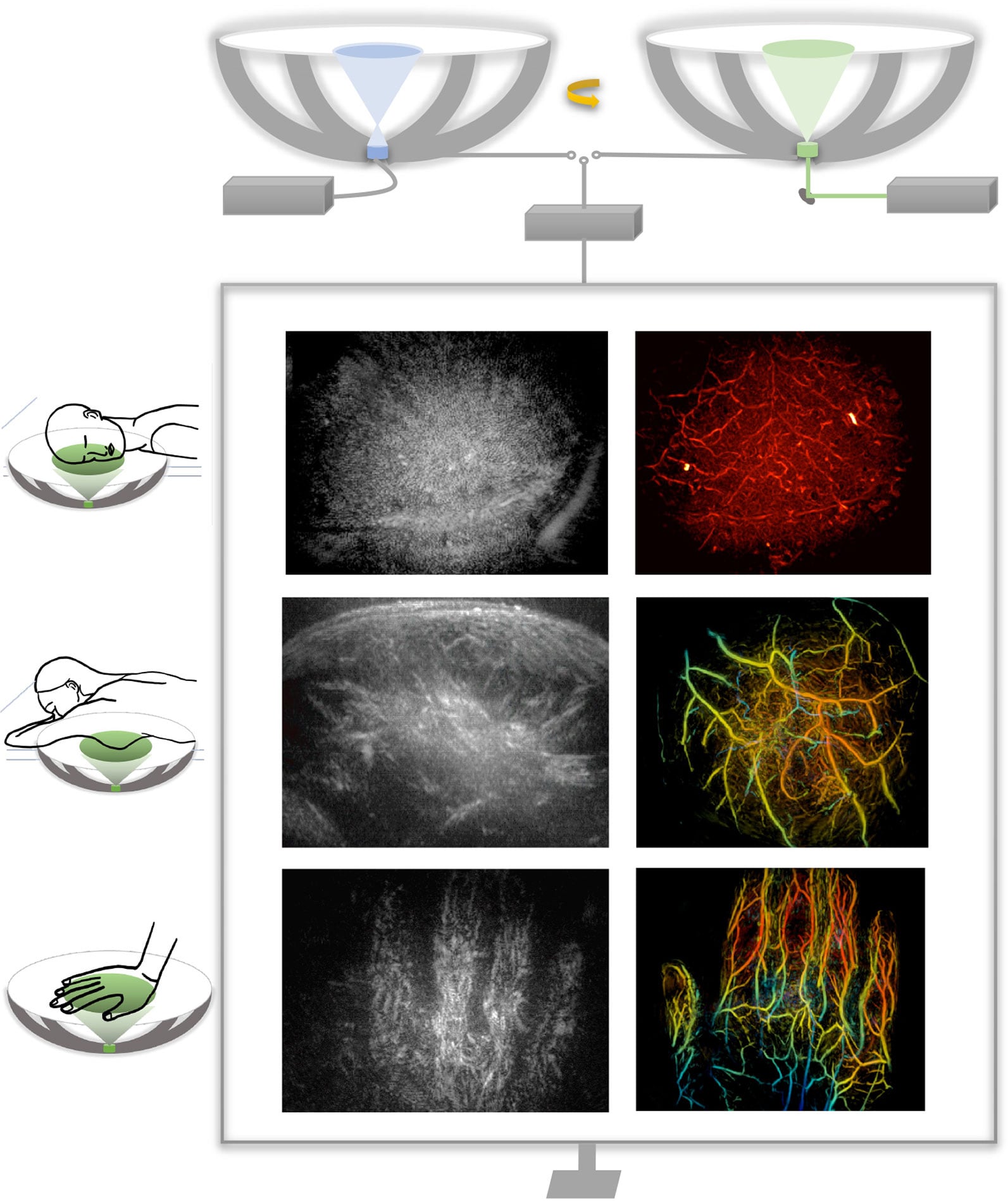
By mixing two highly effective imaging strategies, researchers from Caltech and USC have developed a brand new method to see contained in the human physique with unprecedented pace and element. The method produces three-dimensional, full-color photographs that present not solely the form of soppy tissues but in addition how blood vessels are functioning in actual time. In early demonstrations, the researchers efficiently imaged a number of completely different components of the human physique, highlighting the flexibility of the method.
This mixed imaging technique might considerably enhance how medical doctors detect and research illness. Potential functions embody extra exact breast tumor imaging, new methods to trace nerve harm brought on by diabetes, and superior instruments for observing mind construction alongside blood movement. The work suggests a path towards medical scans which are each extra informative and simpler to repeat over time.
The researchers describe the brand new expertise in a paper printed in Nature Biomedical Engineering.
Medical imaging typically requires tradeoffs between pace, price, and the kind of data that may be captured. Ultrasound, probably the most extensively used strategies, is quick, cheap, and properly suited to visualizing the construction of tissues. Nevertheless, it sometimes offers solely two-dimensional views and can’t simply seize a large space or reveal detailed details about blood chemistry or movement.
Photoacoustic imaging addresses a few of these gaps however introduces others. On this method, laser mild is shipped into the physique, the place it’s absorbed by molecules in blood vessels. That absorption generates sound waves that may be detected and translated into photographs. As a result of completely different molecules take in mild in distinct methods, photoacoustic imaging can show blood vessels in optical colour—permitting for visualization of how blood strikes by way of arteries and veins. By itself, nonetheless, the method doesn’t present sufficient structural element to completely map surrounding tissues.
Different superior imaging instruments, comparable to computed tomography (CT) scanning and magnetic resonance imaging (MRI), can ship detailed views of anatomy, however they arrive with notable downsides. These strategies might be expensive, might require distinction brokers, typically contain publicity to ionizing radiation, or take too lengthy to be sensible for frequent monitoring or bedside use.
Combining Ultrasound and Photoacoustics
To beat these limitations, the researchers developed RUS-PAT (rotational ultrasound tomography, RUST, mixed with photoacoustic tomography, PAT). PAT was first developed greater than 20 years in the past by Lihong Wang, the Bren Professor of Medical Engineering and Electrical Engineering and the Andrew and Peggy Cherng Medical Engineering Management Chair at Caltech.
In PAT, molecules that take in mild reply to brief laser pulses by vibrating, which generates acoustic alerts. These alerts can then be detected and processed to kind detailed, high-resolution photographs.
Wang, who can also be the manager officer for medical engineering at Caltech, says his group’s purpose with the present work was to mix the advantages of PAT with ultrasound. “However it’s not like one plus one,” he says. “We would have liked to seek out an optimum manner of mixing the 2 applied sciences.”
Ultrasound sometimes makes use of many transducers to each generate and obtain ultrasound waves, and mixing this course of straight with PAT could be too advanced and costly for widespread use. PAT, in the meantime, solely requires the detection of ultrasound, and that gave Wang an concept. “I assumed, ‘Wait, can we simply mimic mild excitation of ultrasound waves in photoacoustic tomography, however do it ultrasonically?'” PAT permits laser mild to diffuse inside the tissue, in the end triggering the manufacturing of measurable ultrasound waves. Equally, Wang figured, they may use a single wide-field ultrasound transducer to broadcast an ultrasonic wave broadly into the tissue.
They may then use the identical detectors to measure the ensuing waves for each modalities. Within the new system, a small variety of arc-shaped detectors are rotated round a central level, permitting it to behave like a full hemispheric detector however at a fraction of the complexity and value.
Demonstrated Scientific Potential
“The novel mixture of acoustic and photoacoustic strategies addresses lots of the key limitations of extensively used medical-imaging strategies in present scientific follow, and, importantly, the feasibility for human software has been demonstrated right here in a number of contexts,” says Dr. Charles Y. Liu, an writer of the paper who’s a visiting affiliate in biology and organic engineering at Caltech. Liu can also be a professor on the Keck Faculty of Drugs of USC, director of USC’s Neurorestoration Middle, and chair of neurosurgery on the Rancho Los Amigos Nationwide Rehabilitation Middle.
The RUS-PAT method might doubtlessly be utilized in any area of the physique to which mild might be delivered, and for functions the place clinicians or researchers would profit from the synergistic imaging of each the morphology and color-related operate. For instance, RUS-PAT might enhance breast-tumor imaging, giving physicians the power to know a tumor’s actual location and environment in addition to its pathology and physiology. It might additionally assist medical doctors monitor the nerve harm brought on by diabetic neuropathy by offering an all-in-one method to monitor oxygen provide together with morphology. Wang says the method is also helpful in mind imaging, permitting scientists to watch the structural particulars of the mind whereas additionally having the ability to observe hemodynamics.
At present, the system can scan to a depth of about 4 centimeters. Gentle will also be delivered endoscopically, doubtlessly making deeper tissues accessible to the brand new expertise. A RUS-PAT scan might be carried out in lower than one minute.
The present setup entails a scanning system with ultrasound transducers and laser housed beneath a mattress. It has been demonstrated on human volunteers and sufferers and is within the early levels of translational growth.
Reference: “Rotational ultrasound and photoacoustic tomography of the human physique” by Yang Zhang, Shuai Na, Jonathan J. Russin, Karteekeya Sastry, Li Lin, Junfu Zheng, Yilin Luo, Xin Tong, Yujin An, Peng Hu, Konstantin Maslov, Tze-Woei Tan, Charles Y. Liu and Lihong V. Wang, 16 January 2026, Nature Biomedical Engineering.
DOI: 10.1038/s41551-025-01603-5
The work was supported by funding from the Nationwide Institutes of Well being.
